Light hitting a plant or the human eye triggers complex molecular changes. But molecules that react to light undergo structural changes so fast — in less than 1 trillionth of a second — that the initial stages of photosynthesis and vision remain largely a mystery.
A research group led by Martin Centurion, assistant professor of physics and astronomy, has discovered a way to peer into that unknown molecular world. Their ability to capture three-dimensional images of molecules undergoing structural changes is a major advance toward studying how light energy is converted into chemical energy. The technique may one day lead to better alternative energy sources, help solve vision problems and improve skin cancer prevention and treatment.
To get a 3-D image, molecules must be floating in a gas state. "When you have molecules in a gas, they are rotating," Centurion said. "It's very hard to take a three-dimensional image of it because you cannot hold it."
Previous 3-D structures were based on models made from two-dimensional images. "With our method, you never go to a model. You go from the data to the structure of the molecule, so it's much more powerful in that way," he said.
To gather data, the team first hits the molecules with a laser pulse, which gives them torque, and, for just a split second, the molecules point in the same direction. The laser also triggers a short pulse of electrons that hit the molecules during this brief moment and scatter. By analyzing the electron scatter, Centurion and his team recreate the molecule's structure, like taking its 3-D picture.
So far, they have created an image of a simple five-atom molecule to prove the technique works. The team reported this research in the Sept. 28 issue of the journal Physical Review Letters.
Next, they will use this approach to study molecules that undergo light-induced structural changes. By lengthening the timing between the laser pulse and the electron snapshot, the researchers can create a movie of a molecule as it changes, which will provide insight into its function.
"In principle we can, and plan to, investigate the interaction of molecules with light on a fundamental level, and this can lead to applications across many fields," Centurion said. "We are now getting to the more exciting part of the project."
For example, Centurion's team plans to observe molecules involved in photosynthesis to illuminate how plants convert photons of light into energy. Understanding this process could lead to improving solar energy efficiency and storage. Similarly, observing changes in retinal molecules that absorb light could one day lead to artificial vision technology.
In a biological application, the team will study the pieces of DNA that absorb ultraviolet light. DNA damage from UV light, particularly sunlight, has been linked to skin cancer, though the mechanisms aren't well known. Understanding how light damages DNA could lead to better treatment or protection options.
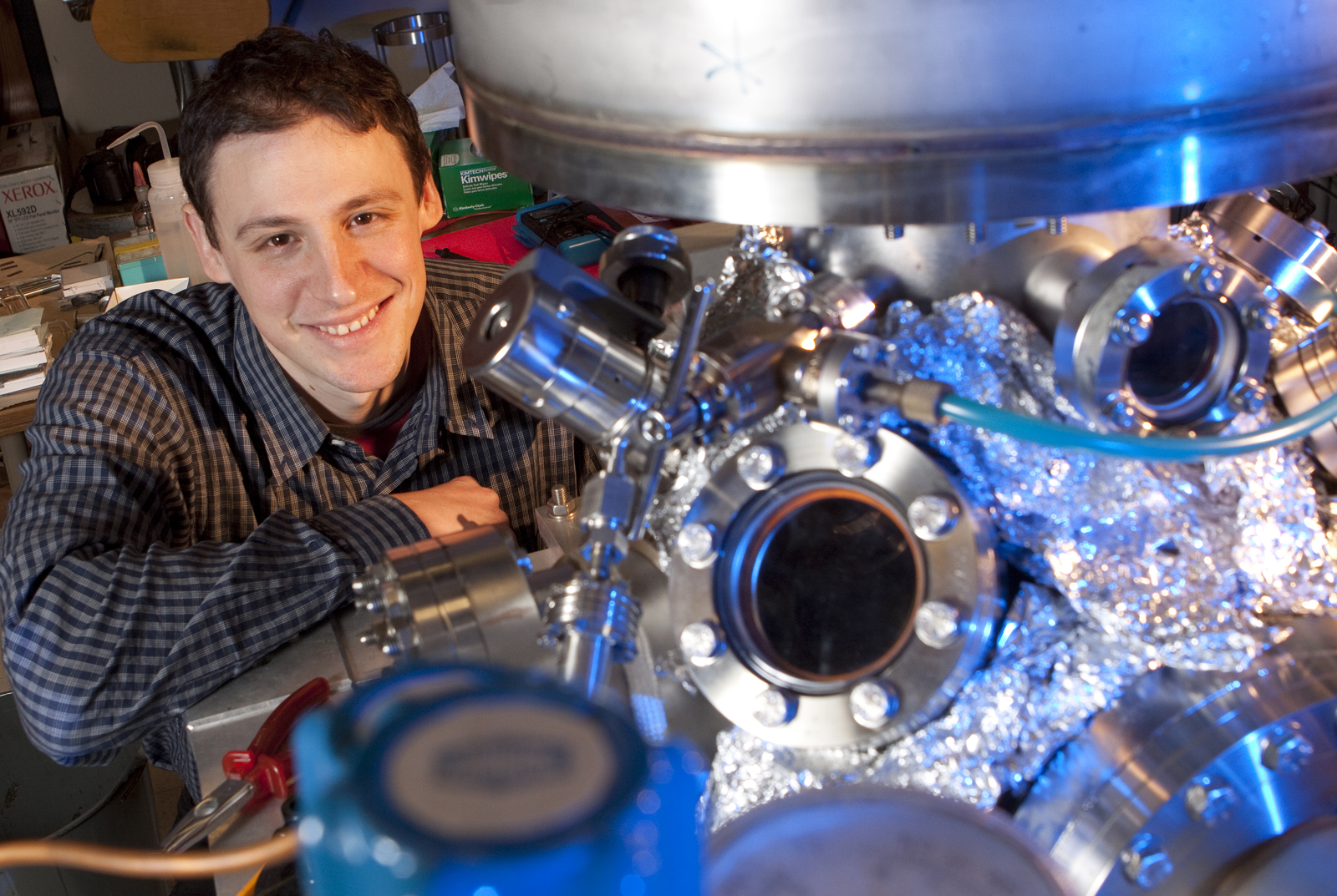
Centurion's team includes graduate student Jie Yang and postdoctoral fellow Chris Hensley. A $750,000 Department of Energy Early Career Research Program award funds this research.
— Gillian Klukas, Research and Economic Development